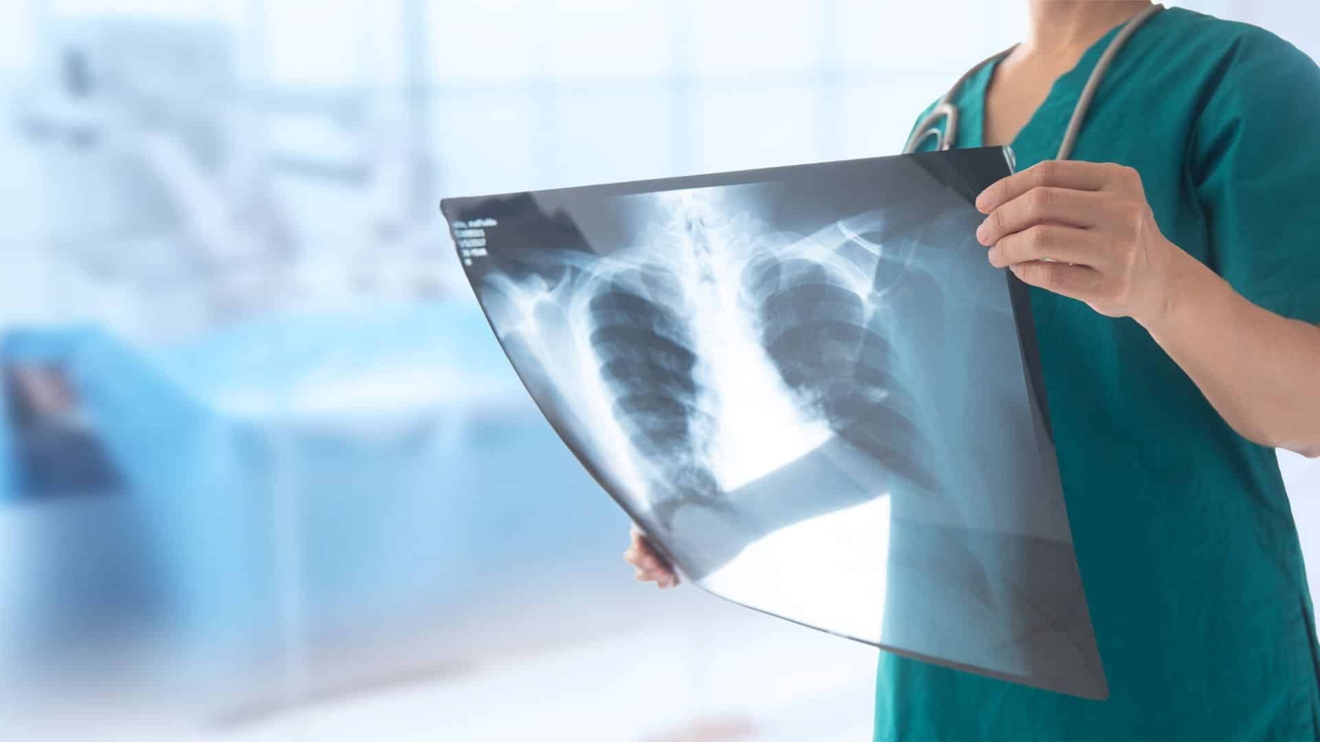
X-ray imaging In the dynamic landscape of medical diagnostics, stands as a cornerstone, providing invaluable insights into the internal structures of the human body. Widely recognized for its role in detecting bone fractures, this powerful imaging technique has evolved over the years to become a crucial tool for diagnosing various medical conditions. Explore with us X-ray imaging, exploring its applications, advantages, and the journey of medical images from the X-ray machine to our ER Physicians.
Unveiling the World of X-ray Imaging
X-ray imaging is a medical procedure that utilizes a small amount of ionizing radiation to create detailed pictures of the inside of the body. This process involves passing X-ray beams through the body, with varying degrees of radiation absorption by different tissues, producing detailed images. The outcome visually represents bones, organs, and other structures, empowering healthcare professionals to identify abnormalities, assess injuries, and make informed diagnostic decisions. (Mayo Clinic, 2022)
When is an X-ray Necessary?
The decision to employ X-ray imaging is often based on the specific diagnostic needs of a patient. X-rays excel at visualizing dense structures such as bones, making them particularly useful for diagnosing fractures, joint dislocations, and bone infections. X-rays also play a pivotal role in detecting lung conditions like pneumonia and lung cancer.
While X-rays provide exceptional imaging of bones, they may be less effective in visualizing soft tissues like muscles and tendons. In such cases, healthcare providers may recommend alternative imaging modalities such as CT scans or MRIs. CT scans offer detailed cross-sectional images, ideal for examining soft tissues and organs. At the same time, MRI utilizes powerful magnets and radio waves to create clear pictures of soft tissues without using ionizing radiation.
The choice between X-ray, CT, or MRI depends on the specific clinical scenario and the information sought by the healthcare provider. X-ray imaging remains a rapid and cost-effective option for many diagnostic needs, mainly when a swift assessment of bone structures is essential. (Johns Hopkins, 2022)
Risks of X-ray
Despite their utility, x-rays emit ionizing radiation, capable of causing harm to living tissue, with risks accumulating over a lifetime. Although the likelihood of developing cancer from radiation exposure is generally low, caution is warranted.
For pregnant women, x-rays present minimal risks to the baby if imaging doesn’t involve the abdomen or pelvis. Preferably, alternative imaging methods like MRI or ultrasound are favored for abdominal and pelvic examinations during pregnancy. In emergencies or time constraints, however, x-rays may be considered. Due to heightened sensitivity and longer life expectancy, children face a relatively higher risk of developing cancer from ionizing radiation. Parents are encouraged to inquire about machine settings adjusted for pediatric use, emphasizing the importance of balancing diagnostic benefits with potential risks in medical imaging decisions.(National Institute of Biomedical Imaging and Bioengineering)
The Journey of Medical Imaging in the U.S.
Once an X-ray is conducted, the image travels from the X-ray machine to the hands of your healthcare provider. After capturing X-ray images, they are stored digitally, enhancing accessibility for healthcare professionals to view and analyze them on computer screens.
The medical images are securely transmitted from digital storage to a picture archiving and communication system (PACS). PACS is a technology that facilitates medical image storage, retrieval, and distribution, providing centralized access for authorized healthcare providers. This system ensures efficient collaboration and timely decision-making.
To further enhance communication between healthcare professionals and patients, many medical institutions in the U.S. offer online portals where individuals can access their medical images and reports. This transparency fosters patient engagement and empowers individuals to participate actively in their healthcare journey. (Radiological Society of North America (RSNA) and American College of Radiology (ACR))
Diagnostic Imaging at Our Facility
X-ray imaging remains a vital tool in medical diagnostics, offering a detailed view of the human body’s inner workings. Its applications extend beyond bone-related conditions, providing valuable insights into various medical issues. Understanding when to opt for X-rays, considering alternative imaging methods, and appreciating the journey of medical images from the X-ray machine to your healthcare provider contributes to a more informed and empowered healthcare experience.
Works Cited
Mayo Clinic. “X-Ray.” Mayo Clinic, Mayo Foundation for Medical Education and Research, 11 Feb. 2022, www.mayoclinic.org/tests-procedures/x-ray/about/pac-20395303.
Johns Hopkins Medicine. “CT Scan versus MRI versus X-Ray: What Type of Imaging Do I Need?” Johns Hopkins Medicine, 6 Sept. 2022, www.hopkinsmedicine.org/health/treatment-tests-and-therapies/ct-vs-mri-vs-xray.
Radiological Society of North America (RSNA) and American College of Radiology (ACR). “How to Obtain and Share Your Medical Images.” Radiologyinfo.Org, www.radiologyinfo.org/en/info/article-your-medical-images.
National Institute of Biomedical Imaging and Bioengineering. “X-Rays.” National Institute of Biomedical Imaging and Bioengineering, U.S. Department of Health and Human Services, www.nibib.nih.gov/science-education/science-topics/x-rays.

