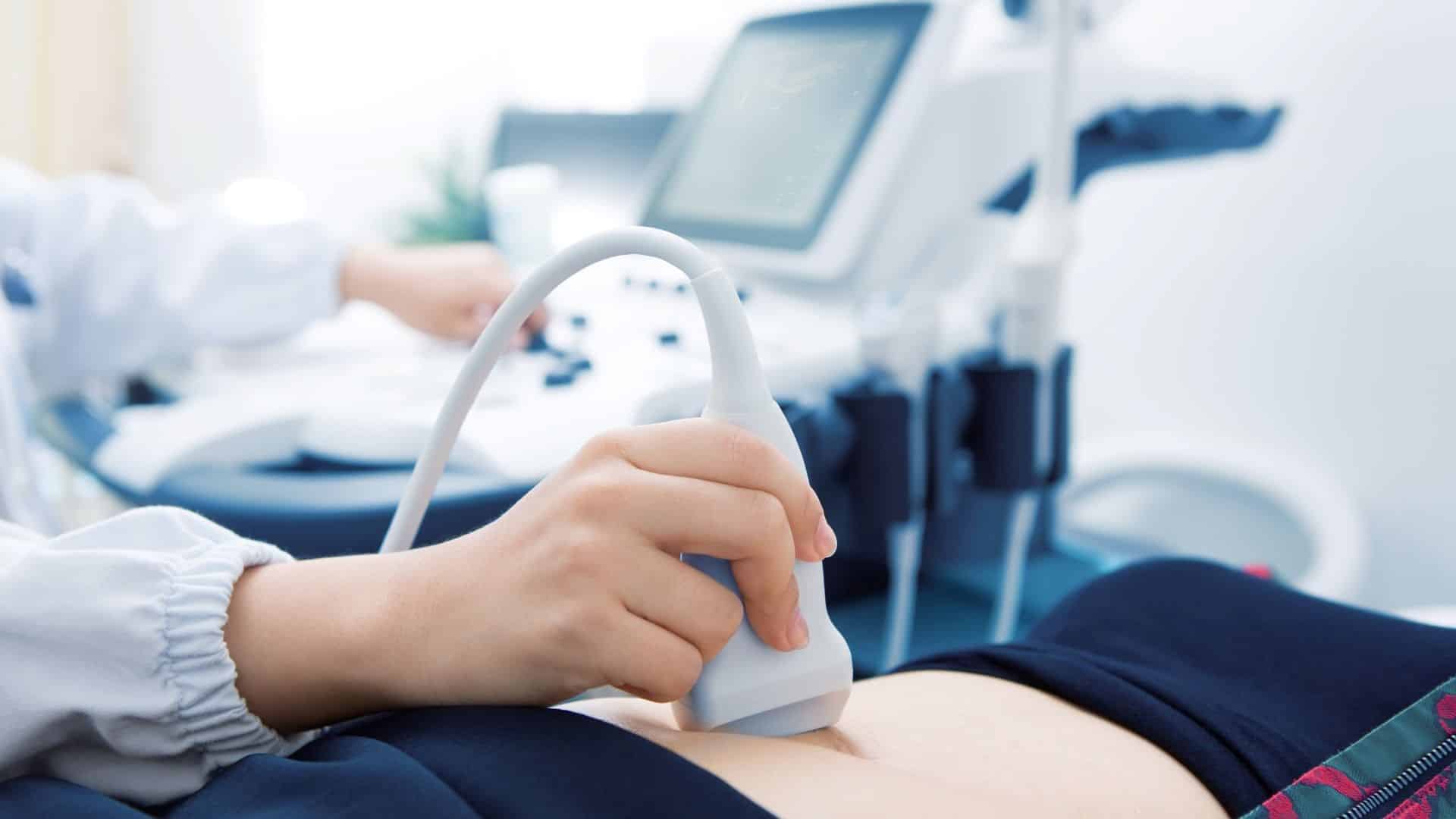
Ultrasound diagnostics, a technological tool, has transformed the way we scan the human body. By harnessing sound waves to produce detailed internal images, ultrasound has become a vital tool across medical specialties. Let’s delve into the world of ultrasound diagnostics, uncovering its applications, advantages, and the technology that powers it.
Understanding Ultrasound
Ultrasound, or sonography, is a non-invasive imaging technique that employs high-frequency sound waves to create real-time images of the body’s internal structures. Unlike X-rays or CT scans, ultrasound does not use ionizing radiation, making it a safer option for patients and healthcare providers. Instead, it relies on the principles of sound wave propagation. It echoes to generate detailed images of organs, tissues, and blood vessels.
The ultrasound process begins with a transducer, a handheld device that emits high-frequency sound waves into the body. These waves travel through the body and bounce back as echoes when encountering different tissues. The transducer captures these echoes, and a computer processes the information to create dynamic images displayed on a monitor in real-time. (WebMD)
Uses of Ultrasound
According to experts, ultrasound is a versatile tool used in various medical fields, including obstetrics, cardiology, gastroenterology, and musculoskeletal imaging. Its ability to visualize soft tissues, such as the liver, kidneys, and reproductive organs, makes it an indispensable diagnostic tool.
In obstetrics, ultrasound is commonly used to monitor the development of a fetus during pregnancy. The safety of ultrasound during pregnancy is unparalleled, as it does not expose the fetus to ionizing radiation. The detailed images obtained through ultrasound assist healthcare providers in assessing fetal growth, detecting abnormalities, and ensuring a healthy pregnancy.
Cardiac ultrasound, or echocardiography, is another critical application. This technique allows healthcare professionals to visualize the heart’s structure and function, helping diagnose heart conditions, assess blood flow, and monitor cardiac health. (Mayo Clinic, 2022)
Diagnosing Abdominal, Pelvic, and Vascular Conditions
Ultrasound is useful in identifying and evaluating conditions affecting the abdomen, pelvis, and blood vessels. The clinic emphasizes the role of ultrasound in diagnosing liver and gallbladder diseases, detecting kidney stones, and evaluating the reproductive organs.
It assesses blood flow and detects potential issues in the blood vessels. This non-invasive method aids in diagnosing conditions like deep vein thrombosis (DVT) and arterial blockages, contributing to timely intervention and improved patient outcomes. (Cleveland Clinic)
Internal & External Ultrasound
Ultrasound examinations are valuable diagnostic tools employed for various medical purposes, distinguished into external and internal procedures. In external ultrasound, a sonographer applies a lubricating gel on the patient’s skin and places a transducer on the lubricated area, moving it over the targeted body part, such as the heart or fetus in the uterus. Discomfort is minimal, with patients primarily sensing the transducer on their skin. In pregnancy, slight discomfort may occur due to a full bladder.
Conversely, internal ultrasound involves assessing internal reproductive or urinary organs. Evaluating digestive organs like the esophagus may require an endoscope, incorporating a light and ultrasound device inserted through the mouth. Medications are often administered before internal procedures to alleviate potential discomfort.
Ultrasound procedures are generally noninvasive and lack ionizing radiation, rendering them safe. However, long-term risks remain uncertain, discouraging unnecessary “keepsake” scans during pregnancy. Medical necessity dictates the recommendation for ultrasound during pregnancy. (Medical News Today)
Versatile Diagnostic and Guidance Tool in Medical Procedures
Ultrasound guidance is commonly used in biopsies, helping healthcare providers precisely target specific areas for tissue sampling.
The dynamic nature of ultrasound imaging showcases its ability to capture real-time movement within the body. This feature is precise in musculoskeletal ultrasound, where healthcare providers can assess joint and soft tissue conditions, such as tendon injuries and inflammation, as the patient moves. (WebMD)
Ultrasound: Diagnostic Excellence
From monitoring fetal development to diagnosing cardiac conditions and guiding medical procedures, ultrasound is a cornerstone in modern healthcare. The non-invasive nature of ultrasound and its real-time imaging capabilities make it a preferred choice for patients and healthcare providers. As we navigate the world of medical diagnostics, ultrasound promises great precision and insight into the human body’s complexities.
Works Cited
WebMD. “Abdominal Ultrasounds: Purpose, Procedure, Uses, Results, Benefits.” WebMD, www.webmd.com/a-to-z-guides/what-is-an-ultrasound.
Mayo Clinic. “Ultrasound.” Mayo Clinic, Mayo Foundation for Medical Education and Research, 30 Apr. 2022, www.mayoclinic.org/tests-procedures/ultrasound/about/pac-20395177.
Cleveland Clinic. “Ultrasound: What It Is, Purpose, Procedure & Results.” Cleveland Clinic, my.clevelandclinic.org/health/diagnostics/4995-ultrasound.
Medical News Today. “Ultrasound Scans: How Do They Work?” Medical News Today, MediLexicon International, www.medicalnewstoday.com/articles/245491#Safety.

